
Overview:
Calcific tendinopathy of the shoulder, sometimes called calcific tendonitis, is when calcium deposits develop inside one or more of the rotator cuff tendons. The supraspinatus tendon is the one most often affected.
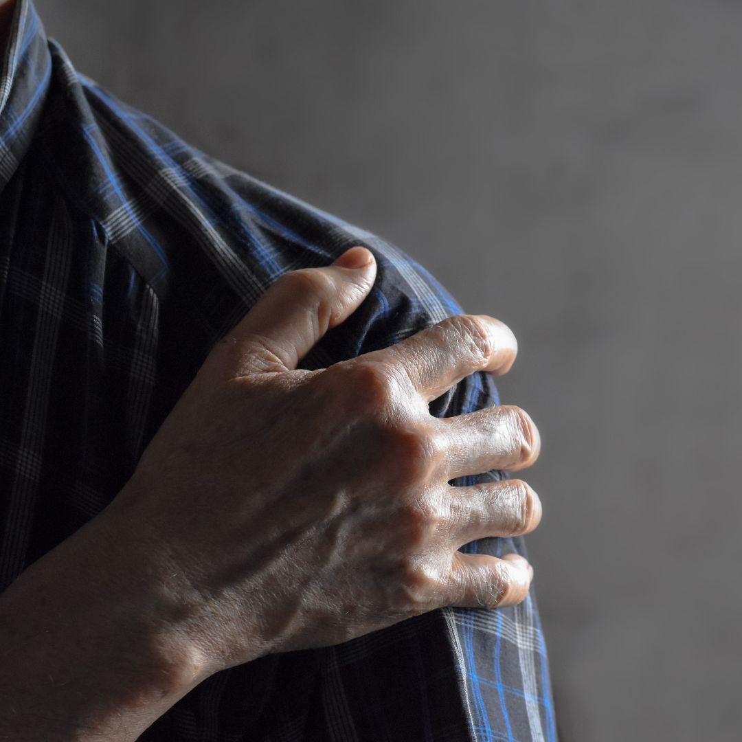
Some people have deposits and no symptoms at all. In others it causes significant shoulder pain, reduced movement, broken sleep and real difficulty lifting or reaching overhead. Symptoms can build slowly or arrive very suddenly. During an acute flare the pain can become intense without any obvious injury, which is one reason people end up in an emergency department wondering what they have done.
Treatment depends on the severity and how long it has been going. Many cases are managed conservatively with activity modification, exercise rehabilitation and symptom management, and shockwave therapy is worth considering for persistent cases. This page sits under our wider shoulder pain information.
Anatomy:
The rotator cuff is four muscles and their tendons: supraspinatus, infraspinatus, teres minor and subscapularis. They surround the shoulder joint, stabilise the head of the humerus in the socket, lift and rotate the arm, and control it through reaching and overhead movement.
Calcific tendinopathy occurs when calcium hydroxyapatite deposits develop within one of those tendons, most commonly the supraspinatus. The subacromial-subdeltoid bursa sitting nearby can also become irritated, and that is often what makes an acute episode so painful.
Why do the deposits form? The exact reason is not fully understood. It is not caused by eating too much calcium, by high dietary calcium levels, by posture alone, or simply by wearing the shoulder out. It appears to be a biological process within the tendon itself.
Causes:
Calcific tendinopathy most commonly affects adults between roughly 30 and 60. Many people develop it with no obvious risk factor at all. Associations that have been described include:
- Age: most cases fall in that 30 to 60 bracket.
- Biological factors affecting tendon tissue: the underlying process is within the tendon rather than mechanical.
- Metabolic conditions: in some individuals.
- Endocrine conditions: in some patients.
- Repetitive shoulder loading: though this is not the whole story and many cases have no loading history.
The stages matter more than the cause. In the formative phase calcium gradually accumulates, and symptoms range from mild and intermittent to none. In the resting phase the deposit sits there, again with anything from no pain to persistent discomfort. In the resorptive phase the body starts breaking the deposit down, and this is usually the most painful stage. The deposit softens and the surrounding tissue becomes highly irritated, which can produce sudden severe pain. In the post-calcific phase the tendon remodels and symptoms typically improve. Not everyone follows that progression neatly.
Symptoms:
- Pain at the front or outer shoulder: the usual location.
- Severe pain with no obvious injury: a hallmark of an acute resorptive episode.
- Pain raising the arm: and during overhead activity.
- Pain reaching behind your back: and difficulty dressing.
- Night pain: lying on that side, often disturbing sleep badly during a flare.
- Reduced movement and weakness: usually because of pain rather than true loss of strength.
- Difficulty lifting, training or doing physical work: which is often what brings people in.
During an acute resorptive episode, pain can be severe enough to significantly restrict shoulder movement and interfere with sleep. That intensity is not a sign of permanent damage.
Diagnosis:
Assessment covers the same ground as any shoulder presentation, with one difference: because calcium deposits show up clearly on imaging, scans are more relevant here than for most uncomplicated shoulder pain.
- Location and behaviour of the pain: what provokes it, what settles it, and how quickly it came on.
- Shoulder range of motion: and how much of the restriction is pain rather than stiffness.
- Rotator cuff strength: tested individually.
- Pain on resisted movement: which directions reproduce your symptoms.
- Shoulder blade function: how the scapula moves on the ribcage.
- Work, gym and sporting demands: and any previous shoulder problems.
- Imaging: X-ray often shows the deposit clearly, including its location, size and appearance. Ultrasound also identifies deposits and assesses the cuff tendons and the bursa, and can guide procedures where needed. MRI is not usually required just to find calcification, but may be recommended where other significant shoulder pathology is suspected.
Calcific tendinopathy or rotator cuff tendinopathy? Both involve the cuff and produce similar symptoms. The difference is the calcium deposit. You can also have both together, or calcific tendinopathy alongside shoulder bursitis, or calcium on a scan with no shoulder pain at all. Finding calcium does not automatically prove it is responsible for everything you are feeling. Our rotator cuff tendinopathy page covers the non-calcific picture.
Treatment:
Treatment depends on the severity of symptoms, the stage, the size and character of the deposit, how long it has been going, the functional limitation and what has already been tried. Most cases can be managed without surgery.
- Activity modification: during painful periods, temporarily reducing heavy overhead lifting, repetitive reaching, high-volume swimming, throwing, heavy pressing and the specific gym exercises that hurt. Complete long-term rest is not the objective, and activity is rebuilt as symptoms settle.
- Exercise rehabilitation: progressive work through rotator cuff strength, deltoid strength, the scapular muscles, shoulder endurance and functional lifting capacity. During a severe flare this starts gentle and builds as pain becomes manageable.
- Shoulder mobility: significant pain makes people stop moving the shoulder. Mobility work maintains or restores movement while symptoms settle, which matters most where pain has caused prolonged avoidance.
- Manual therapy: useful for associated muscular or joint stiffness and short-term relief. It does not remove calcium deposits, so it sits alongside active rehabilitation.
- Shockwave therapy: extracorporeal shockwave delivers controlled acoustic waves to the area and is worth considering for persistent calcific rotator cuff tendinopathy. It is available at every Tensegrity clinic except Willoughby, and is used alongside a rehabilitation program rather than instead of strengthening and progressive loading. Not everyone with calcium on a scan needs it. That decision comes from your symptoms and examination, not the image.
- Review and progression: reassessed against function as the stage changes.
Pain-relieving or anti-inflammatory medication may be appropriate during painful episodes and is worth discussing with your GP or pharmacist, particularly when symptoms are severe. A corticosteroid injection is sometimes considered where associated bursal pain is limiting function, though it does not remove the deposit. For selected persistent cases, ultrasound-guided barbotage, where a needle is guided to the deposit and it is broken up, irrigated or partially aspirated, may be considered. That is performed by an appropriately trained medical practitioner or radiologist and is not needed in every case. Most people do not require surgery.
Rehabilitation:
How long does it take? Recovery varies a lot, because the condition moves through different biological stages. An acute painful episode often improves substantially over several weeks. More persistent cases can need around two to six months or longer of management and rehabilitation.
Some deposits stay visible on imaging even after symptoms have improved, and changes in the deposit do not always bring immediate pain relief. So recovery is judged on pain, movement, strength, sleep, function, and return to work and sport, rather than on the scan.
Getting back to the gym and sport. Progressive rather than all at once. Painful overhead movements are modified first, then rehabilitation moves through comfortable movement, basic cuff strengthening, progressive resistance, overhead strengthening, heavier pushing and pulling, faster movement, sport-specific work and finally normal volume. For athletes the endpoint is the actual demand of the sport, not the point where everyday pain settles.
Three exercises for calcific tendinopathy of the shoulder
These three were put together by our practitioners as a starting point. They are general exercises, not a personalised program.
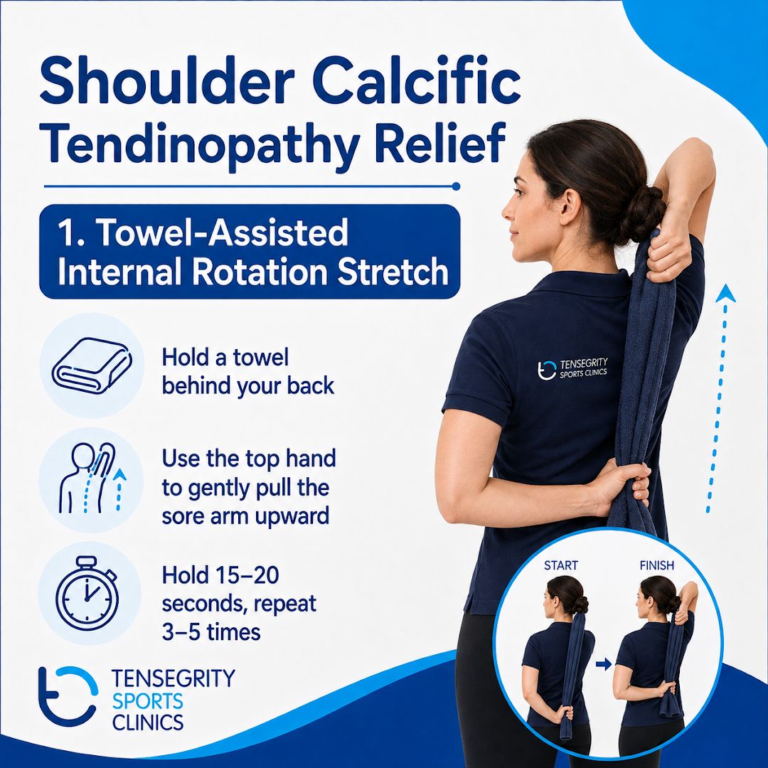
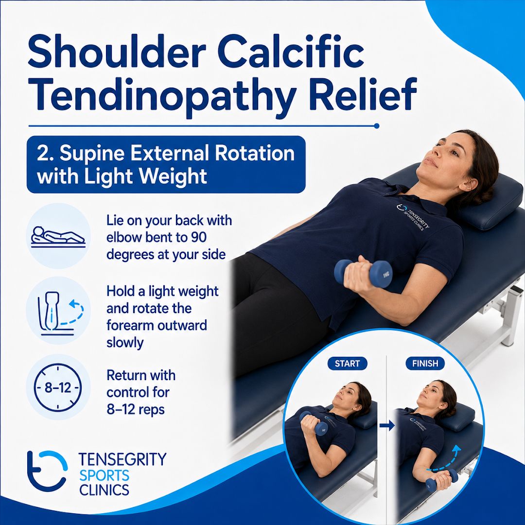
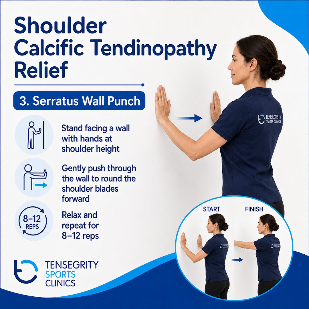
During a severe acute flare these may be too much. Start where you are comfortable and book an assessment so the loading matches the stage you are in.
Prevention:
Because the cause of calcium formation is not fully understood, calcific tendinopathy cannot always be prevented. Keeping the shoulder strong and capable does reduce the impact of an episode.
- Maintain rotator cuff strength: ongoing, not only when something hurts.
- Maintain shoulder blade and upper back strength: so the joint is not carrying everything.
- Progress gym loads gradually: and increase overhead training in steps.
- Allow recovery between demanding sessions: rather than stacking them.
- Keep generally active: regular physical activity supports tendon health.
- Address recurring shoulder symptoms early: before they start limiting you.
- Keep strengthening after the pain improves: which is when most people stop.
Outlook:
The outlook is generally good. The condition can be extremely painful, particularly during the resorptive phase, but severe pain does not mean significant permanent damage has occurred. Many cases improve with time and conservative management. Where symptoms persist, shockwave therapy, image-guided procedures or onward referral for medical assessment may be considered.
Finding calcium in the shoulder sounds alarming and often is not. A deposit does not mean the tendon is permanently damaged. Some eventually reduce or resolve, others stay visible without causing symptoms. The aim is not a normal-looking scan. It is comfortable movement, strength, sleep, function and getting back to what you normally do.
Get assessed if you have: severe shoulder pain with no obvious cause, significant loss of movement, persistent night pain, progressive weakness, symptoms that are not improving, difficulty doing your normal work or daily activities, or significant shoulder pain after trauma.
Seek prompt medical assessment if the shoulder becomes red, hot and significantly swollen, particularly with a fever or feeling generally unwell. That can indicate infection or another condition needing urgent attention.